
The Unexpected Role of Nurse Shark Pancreas as a Secondary Lymphoid Organ
Introduction
The adaptive immune system is compartmentalized into primary and secondary lymphoid organs. In mammals, primary lymphoid organs include the thymus and the bone marrow where immature T cells and B cells develop, respectively. In both tissues, developing lymphocytes undergo RAG-mediated somatic recombination of their antigen receptors followed by several tolerance checkpoints which prevent reactivity to self-antigens. Given the enormous diversity of B cell receptors (BCRs) and T cell receptors (TCRs) generated by the recombination process, the frequency of lymphocytes able to respond to a specific antigen is very low.1 Secondary lymphoid organs (SLOs) provide a structured environment to facilitate interactions between low frequency antigen-specific B and T cells as well as cognate antigen displayed by antigen presenting cells (APCs), resulting in the initiation of effective adaptive immune responses. The spleen is the most evolutionary ancient SLO, being present in all jawed vertebrate lineages.2,3 Generally, splenic cellular organization increases in complexity from the ancient jawed vertebrate lineages, such as cartilaginous fishes, to more recently diverged lineages such as mammals. However, in almost all lineages the spleen contains defined white pulp, where immune cells are primarily located, and red pulp which filters dead and dying red blood cells and helps transport antigens to the white pulp. Embedded within the white pulp are B cell follicles which are bordered by organized T cell zones (eg, mammals) or scattered T cell clusters (eg, cartilaginous fishes).3,4 It is within these B cell follicles that antigen accumulates and is displayed in an intact form, thus maintaining structural epitopes for antigen-specific B cell selection.
In mammals, the spleen coordinates with a complex network of draining lymph nodes and Peyer’s patches of the gut to ensure timely and efficacious adaptive immune responses. The microanatomy of these additional SLOs mirrors that of the spleen; however, the source of antigen surveilled by each SLO varies. While the spleen concentrates antigens directly from the bloodstream, the lymph nodes orchestrate responses to antigens acquired from peripheral tissues via the afferent lymph, and Peyer’s patches sample gut-derived antigens.5 Thus, the presence of diverse SLOs allows the adaptive immune system to employ a “zonal defense” strategy, ensuring that pathogens that infect the peripheral tissues can be detected and appropriate antibody responses initiated before the infection becomes systemic. However, lymph nodes are only present in mammals and possibly some birds,2,3,6 and the GALT found in other vertebrate lineages thus far lacks the cellular organization of true SLOs.3,7 Thus, how the majority of vertebrate species achieve comprehensive immune surveillance of their tissues without a network of extrasplenic SLOs is currently unclear.
To address this question, we investigated the immune tissues and adaptive immune response of the nurse shark (Ginglymostoma cirratum), a cartilaginous fish and model organism for understanding immune system evolution. Cartilaginous fishes (Chondrichthyes; sharks, skates, rays, and chimeras) last shared a common ancestor with other jawed vertebrates approximately 450 million years ago8,9 and are the most ancient extant class to possess an adaptive immune system built upon immunoglobulin (Ig)-based rearranging receptors.10 As in other jawed vertebrate species, shark T cells mature in the thymus.11,12 However, lacking bone marrow, cartilaginous fish B cell maturation occurs in the epigonal organ, a tissue associated with the gonad, and the Leydig organ embedded within the walls of the esophagus in some species.11,13 Thus far, the only verified cartilaginous fish SLO is the spleen, which features defined white pulp comprised of well-organized B cell follicles.2,13,14 These splenic follicles have recently been shown to serve as sites of B cell selection, sharing the functional hallmarks that define mammalian germinal center (GC)-mediated adaptive immune responses, ie, the display of intact antigen acquired from peripheral sites by an unknown APC type, the segregation of activated B cells performing antigen selection from those that are proliferating/mutating their BCRs, and the provision of help, in the form of survival signals, by specialized T cells (specifically Tfh-like cells).14,15 Indeed, long term in vivo studies performed in nurse shark have demonstrated that these processes result in robust, antigen-specific circulating antibody responses which affinity mature and provide lasting immunological memory.16–18
In addition to the spleen, several other shark organs show high levels of secretory Ig, TCR, and other immune gene transcripts.19 However, whether these organs are sites where mature, naïve lymphocytes come into contact with antigen to initiate an adaptive immune response (ie, a SLO) or are simply sites where antigen-experienced lymphocytes reside and/or perform effector functions has not been rigorously evaluated. Here, we investigate shark pancreas as a site of immune activity. High levels of Ig heavy chain transcript have been reported in the pancreas of several shark species, alongside the cytokine B cell activating factor (BAFF) and the BCR-associated signaling molecule CD79a.19–22 Indeed, it has been proposed that the shark pancreas may function as a site of Ig production and aid mucosal protection by secreting antibody directly into the shark intestine via the pancreatic duct.19,23 Using a combination of RNAscope fluorescence in situ hybridization and immunofluorescence microscopy (IF) we discovered that the pancreas contains B cell follicles which are spatially distinct from the exocrine and endocrine pancreas. Further, these “pancreatic B cell follicles” exhibit hallmarks of B cell selection previously identified in nurse shark splenic B cell follicles, and antigen-specific Ig is produced within the pancreas following immunization. Together, our data support the designation of shark pancreas as a SLO and provides insight into how adaptive immune surveillance of the whole organism may function in the absence of a complex lymphatic system.
Materials and methods
Animal maintenance, immunizations, and sample collection
Juvenile nurse sharks (Ginglymostoma cirratum) were acquired from Florida coastal waters under a Special Activity License granted by the Florida Fish and Wildlife Conservation Commission. Animals were transported to the Institute of Marine and Environmental Technology (IMET), Baltimore, where they were acclimatized to captivity in 12,000 l indoor tanks that feature constant recirculating artificial sea water maintained at approx. 28°C. An unimmunized shark (female) was monitored for 1 week following receipt to ensure it was in good health before euthanization. All other animals were held for a minimum of 3 mo to ensure they were in good health prior to immunization. All experiments were performed in accordance with University of Maryland, School of Medicine Institutional Animal Care and Use Committee (IACUC) approved protocols.
Two sharks (1 male and 1 female) were primed subcutaneously in the underside of the pectoral fin with 250 µg R-phycoerythrin (PE) alone or PE plus recombinant severe acute respiratory syndrome coronavirus 2 (SARS2) spike protein displayed upon ferritin nanoparticles (SpFn)24 emulsified in Freund’s complete adjuvant. These animals were boosted intravenously with soluble PE 10 d prior to euthanization on day 40 (PE alone) or day 50 (PE+ SARS2 SpFn).
The third animal (male) was primed subcutaneously in the pectoral fin with 300 µg of ferritin nanoparticles (Fn) emulsified in Freund’s complete adjuvant then boosted 30 d later with 300 ug Fn emulsified in Freund’s incomplete adjuvant, delivered subcutaneously to the opposite pectoral fin. At 60 d this animal was boosted a second time with 300 µg of soluble Fn mixed with 200 µg of PE delivered intravenously (no adjuvant). At 90 d this animal received a final intravenous boost of 200 µg soluble PE and was euthanized 10 days later (ie, 100 d post-prime).
Samples were also utilized from three additional nurse sharks (all male) obtained during a previous study.14 These animals were primed with PE/Freund’s complete adjuvant then euthanized either 40- or 50-d post-priming.
All animals were euthanized with an overdose of tricaine methanesulfonate (MS-222). Following euthanasia, nurse shark tissues were harvested, embedded in Tissue-Tek optimum cutting temperature (OCT) compound (Sakura), and flash frozen in liquid nitrogen. Tissue samples were also taken for RNA preparation; these were diced, placed into cryotubes, and flash frozen in liquid nitrogen. All tissue samples were stored at −80°C.
Immunofluorescence microscopy
OCT-embedded tissues were sectioned on a Tissue-Tek Cryo3 cryostat (Sakura) at 6 µm thickness then adhered to Superfrost Plus microscope slides (VWR). Tissue sections were fixed in ice cold acetone for 30 sec, then rehydrated in PBS for 5 min. The slides were then air dried, and a hydrophobic barrier drawn around each tissue section with a PAP pen. To permeabilize the tissues, each slide was immersed in 0.05% Tween20 in PBS (PBST) for 30 min at 4°C. Tissue sections were then blocked with 10% fetal bovine serum (FBS) in PBST for 45 min at 4°C. Each slide then was incubated with anti-nurse shark IgNAR or anti-nurse shark Ig kappa light chain (as a proxy for IgM) monoclonal supernatants. For staining of IgW, a rabbit polyclonal antibody (IgWv1) previously verified to bind the V-region of recombinant IgW,25 was used. Isotype controls corresponding to each primary antibody were included as negative controls. Following incubation with primary antibodies, slides were washed 3 times with PBST for 5 min. Slides were then incubated with goat anti-mouse IgG-Alexa Fluor 488 or goat anti-rabbit IgG-Alexa Fluor 488 (Sigma Aldrich) 1:250 in 2% FBS in PBST in the dark for 1 h at 4°C. Slides were washed 3 times with PBST for 5 min, followed by a final rinse in PBS. Slides were air-dried and mounted with ProLong Gold + DAPI (ThermoFisher Scientific). Immunofluorescence microscopy images were taken on an Echo Revolve fluorescence microscope equipped with DAPI, FITC, and TexasRed filters. Images were edited for brightness and contrast using ImageJ.
RNAscope in situ hybridization
RNAscope in situ hybridization was performed using Advanced Cell Diagnostics (ACD) RNAscope Multiplex Fluorescent V2 Assay on frozen OCT embedded tissues sectioned at 6 µm thickness. RNAscope probes were designed and produced by ACD against the nurse shark sequences for CD79a, CD3e, AID, Ki67, TCR-γ, TCR-α, IgM heavy chain, IgNAR, IgW heavy chain, CXCR4, and CXCR5. Probes targeting amylase (XM_048601958.1) and insulin (XM_020521289.2) were generated using sequences mined from the published whale shark (Rhincodon typus, a close relative of nurse shark) genome (Table S2). Assays were conducted following the manufacturers provided protocols. In brief, tissue sections were fixed in 10% neutral buffered formalin for 15 min, rinsed in PBS, then dehydrated across a series of ethanol washes (50%, 75%, and 100%) for 5 min each. Slides were then air dried for 5 min and hydrophobic borders drawn around each tissue section with a PAP pen. Slides were treated with hydrogen peroxide for 10 min then the manufacturers provided protease IV for 30 min. Probes were hybridized to each slide for 2 h at 40°C within a humidified chamber. Negative control probes supplied by ACD and targeting the bacterial DapB gene were used to assess background binding. Probes were amplified with ACD reagents for their specific amplification channel. Opal 520 and Opal 620 fluorophores were used at a 1:1000 dilution to ensure optimal fluorescent signal for each probe. Probed tissue sections were mounted in ProLong Gold + DAPI and fluorescent microscopy images taken on an Echo Revolve fluorescence microscope. Images were edited for brightness and contrast using ImageJ.
Gene expression analysis
Prior to gene expression analysis a series of tissues (pancreas, spleen, epigonal, spiral valve, gill, kidney, brain) were harvested from 3 adult nurse sharks 50 d following immunization with PE and human serum albumin. Tissues were diced into small pieces and placed in cryotubes, flash frozen in liquid nitrogen, and stored at −80°C. Total RNA was extracted from each tissue using the RNeasy kit from Qiagen, following the manufacturer’s instructions and including the DNase I treatment step to remove any contaminating genomic DNA. Purified RNA was quantified using a Qubit 3.0 Fluorometer and stored at −80°C until use. Prior to gene expression analysis 1 µg of RNA from each tissue was converted to cDNA using the GoScript™ Reverse Transcriptase kit (Promega) following provided instructions. No reverse transcriptase controls were also included in this step.
cDNA from each tissue was used in reverse transcription quantitative polymerase chain reaction (RT-qPCR) analysis. RT-qPCR reactions were set up as follows; 10 µl of Power SYBR™ Green PCR Master Mix (Invitrogen), 5 µl of cDNA, 1 µl of forward primer, 1 µl of reverse primer, and 3 µl of DNase/Rnase free water. Reactions were run on a QuantStudio 3 Real Time PCR Machine (ThermoFisher Scientific). Primers were designed against the sequences for nurse shark CD79a and CD3e. Primers targeting insulin (XM_020521289.2) and amylase (XM_048601958.1) were designed against whale shark sequences (Table S1). All primers were validated using melt curve analysis and their efficiencies calculated using a standard curve. Each qPCR reaction was run in duplicate, with “no template” and “no reverse transcriptase” reactions included as controls. To calculate the relative expression of each transcript, CT values were normalized to the combined expression of reference genes RPS13 and RPS29, as these were shown to be stably expressed across our nurse shark tissue panel using the geNORM algorithm. Relative expression values were calculated using the 2-ΔΔCt method and assigning a value of 1 to expression of each transcript in brain.
Detection of antigen-specific IgM by enzyme-linked immunosorbent assays (ELISA)
Nurse shark pancreas and spleen tissue lysates were prepared by homogenizing tissue in 1 ml of ice-cold RIPA buffer containing protease inhibitors for 15 min. Tissue homogenates were then centrifuged at 13,000 RPM at 4°C for 10 min, and the supernatants containing the protein lysates removed. The protein in each lysate was quantified using the Pierce™ BCA Protein Assay Kit (ThermoFisher Scientific). Total protein concentrations were normalized between unimmunized and immunized animals to allow comparison.
To detect antigen specific IgM in these pancreas and spleen tissue lysates, 96 well microtiter plates were coated with recombinant severe acute respiratory syndrome coronavirus 2 (SARS-CoV-2) receptor binding domain (RBD) protein, ferritin nanoparticles (Fn), or 5% milk powder in PBS (MPBS) and left to incubate overnight at 4°C. The following day, plates were washed once with 200 µl per well of PBS, then blocked with 200 µl of MPBS for 1 h and washed again once with PBS. Pancreas and spleen tissue lysates were serially diluted 2-fold and 100 µl of each dilution applied to the RBD-, Fn-, and MPBS-coated wells in duplicate. Tissue lysates were left to incubate for 1 h at room temperature. After incubation, plates were washed three times with PBST, then the anti-nurse shark Ig light chain antibody LK10 (Absolute Antibody Ltd) diluted 1:2500 in MPBS was applied to each well to detect bound IgM and incubated for 1 h. Again, plates were washed 3 times with PBST and then 100 µl of a goat anti-mouse IgG HRP secondary antibody (Invitrogen), diluted 1:5000 in MPBS, was applied to each well and incubated for 1 h. Following incubation, the wells were washed 4 times with PBST and 100 µl of TMB liquid substrate was added to each well. TMB was left on the plates for 5 min to develop signal before quenching with 100 µl of 1M sulfuric acid. The absorbance of each well was read at 450 nm on a Molecular Devices SpectraMax M5 microplate reader.
Immunoprecipitation of nurse shark Igs from biotinylated tissue lysate
Nurse shark pancreas tissue lysate was prepared as described above then biotinylated using the EZ-Link™ Sulfo-NHS-Biotin (ThermoFisher Scientific) following the manufacturer’s instructions. Biotinylated lysate was then incubated overnight at 4 °C with the following antibodies targeting each nurse shark Ig isotype: CB5 (anti-IgM monoclonal supernatant), NARV (anti-IgNAR monoclonal supernatant), IgWv1 (anti-IgW polyclonal antibody). Reaction volumes were topped up to 1 ml with NET-NB buffer. Biotinylated lysate was also incubated with isotype controls corresponding to each primary antibody and with NET-NB buffer alone. The following day, protein G Sepharose was added to each tube and incubated for 1 h. The sepharose-antibody complexes were pelleted by centrifugation, washed 10 times through the repeated addition of NET-N buffer, mixing, pelleting by centrifugation, and removal of the wash buffer. The final wash was left to incubate for 1 h before pelleting. Samples were then resuspended in 2x Laemmli sample buffer (BioRad) containing 2-mercaptoethanol and boiled for 10 min. Boiled samples were loaded onto a 1.5 mm thick 4% to 12% NuPAGE Bis-Tris gel (Invitrogen) and run at 100 V for 1.5 h. The NuPAGE gels were transferred onto PVDF membrane using the Trans-Blot Turbo Transfer System (BioRad). The membranes were then blocked in 5% bovine serum albumin (BSA) in PBS for 1 h. Following blocking, membranes were incubated for 1 h with streptavidin-HRP (Sigma-Aldrich) diluted 1:50,000 in 5% BSA. After incubation, membranes were washed three times with PBST for 5 min each. Membranes were developed with liquid TMB blotting substrate (Invitrogen).
Results
The nurse shark pancreas contains organized lymphoid tissue
The nurse shark pancreas is located near the top of the spiral valve, the shark intestine equivalent, near the head of the spleen (Fig. 1A). While the pancreas is attached to the spiral valve by a small duct, dissection proves that it is a discrete, encapsulated organ with a distinctive leaf-shaped morphology and pale pink coloration (Fig. 1A). To confirm our identification of this organ as pancreas we screened for the presence of insulin and amylase transcripts by RT-qPCR. While all other organs tested, including the spleen and spiral valve, showed no detectable insulin (an endocrine hormone) or amylase (an exocrine digestive enzyme) transcript, the pancreas showed extremely high transcript levels for both genes, confirming the identification of this organ as nurse shark pancreas and proving it has both endocrine and exocrine functions (Fig. 1B).
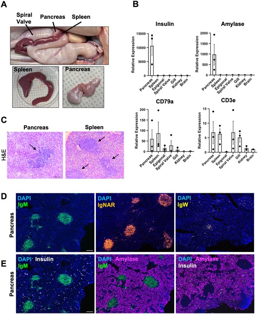
Figure 1.
The nurse shark pancreas contains organized immune tissue. (A) Image showing the anatomical location of the nurse shark pancreas with respect to the spiral valve and spleen. Dissection shows the pancreas is a discrete organ, separate from the spleen, with a distinct morphology and color. (B) RT-qPCR analysis of nurse shark insulin, amylase, CD79a, and CD3e transcripts in select tissues taken from immunized animals. Transcript levels are expressed as arbitrary units normalized against the reference genes RPS13 and RPS29. All RT-qPCR reactions were performed in duplicate wells. The results are presented as the mean ± SE for 3 animals providing 3 biological replicates (n = 3). (C) Hematoxylin and eosin staining of nurse shark pancreas tissue sections identifies dense aggregates of cells (indicated with arrows) visually similar to the B cell follicles found in nurse shark spleen. (D) Representative RNAscope in situ hybridization images of nurse shark pancreas showing the transcript expression of the three nurse shark heavy chain isotypes (IgM, IgNAR, and IgW). (E) Representative RNAscope in situ hybridization images demonstrating the location of IgM+ pancreatic B cell follicles (green) with respect to insulin (white) and amylase (magenta) transcribing cells. (C–E) Images are representative of two nurse sharks that were euthanized 50 d post-prime with PE, providing two biological replicates (n = 2). Scale bars = 100 µm.
Next, to verify the presence of lymphocytes in nurse shark pancreas we used RT-qPCR to screen for transcripts of the confirmed nurse shark B cell marker CD79a and T cell marker CD3e.14 We detected both CD79a and CD3e transcripts in the pancreas at levels comparable to those found in nurse shark spleen, a proven SLO (ie site where B cell responses are initiated),14 supporting the presence of B and T cells within the pancreas (Fig. 1B). Our data align with previous reports of Ig heavy chain and TCR transcripts in the nurse shark pancreas.19,26 Histological examination of pancreas tissue sections revealed the presence of large aggregates of cells, visually similar to B cell follicles found in the white pulp of the nurse shark spleen (Fig. 1C). To ascertain if these cellular aggregates contained B cells, we used RNAscope in situ hybridization on pancreas tissue sections with probes targeting the constant regions of each nurse shark Ig heavy chain. Sharks possess three heavy chain isotypes: IgM, which is produced as a pentamer in a seemingly T cell-independent manner and as a T-dependent monomer, IgNAR a T-dependent heavy-chain only isotype, and IgW, the shark IgD ortholog and proposed shark mucosal isotype.16,19,27 Signal was observed for all 3 Ig heavy chains in the pancreas of immunized animals, each being localized within the observed cellular aggregates (Fig. 1D) in a pattern similar to that observed in the splenic B cell follicles (Fig. S1.1A). Using consecutive sectioning, we found that the heavy chain transcripts of all three Ig isotypes were present in the same “pancreatic B cell follicle,” indicating B cells of different isotypes can populate each follicle similar to the spleen (Fig. S1.1A, B). Importantly, pancreatic B cell follicles are present regardless of immunization status, also being observed in a healthy, unimmunized shark by RNAscope in situ hybridization and immunofluorescent microscopy (Fig. S1.2).
Of note, in one of our immunized animals we observed differential transcription of IgW between individual follicles in both the pancreas and spleen, resulting in “IgW high” and “IgW low” follicles (Fig. S1.3A). This difference was much greater for IgW than IgM and IgNAR (Fig. S1.3B, C). Interestingly, a previous study showed that IgW transcript levels in bulk nurse shark spleen varied substantially between individual animals, possibly as a consequence of their immune state at the time of analysis.19 Our data suggest this may be due to differences in IgW B cell clonal expansion, occurring both at the follicular and organ level.
Given that the existence of pancreatic B cell follicles was wholly unexpected, we next wanted to ensure that the detected Ig heavy chain mRNA transcripts are actively translated into proteins in the pancreas. Using antibodies targeting each Ig heavy chain isotype for IF we confirmed the presence of pancreatic B cell follicles in six different nurse sharks (Fig. S1.4A). Furthermore, the staining intensities for each shark heavy chain isotype were similar to those obtained via RNAscope (Fig. 1D), with IgM+ signal being strongest at both the transcript and protein level, then IgNAR (Fig S1.4B). IgW+ cells were detected much less frequently than cells expressing the other heavy chain isotypes. Notably, in both our IF and in situ hybridization experiments, IgW signal was the lowest of the 3 Ig isotypes in both the pancreas and spleen.
Finally, to determine if the pancreatic B cell follicles are associated with the exocrine or endocrine pancreas, we performed RNAscope in situ hybridization on pancreas tissue sections with probes targeting the transcripts for shark insulin (endocrine hormone), amylase (exocrine enzyme), and IgM heavy chain. Imaging these transcripts demonstrated that the pancreatic B cell follicles are spatially distinct from both amylase- and insulin-expressing cells, suggesting they are not associated with either exocrine or endocrine pancreas (Fig. 1E.). Interestingly, insulin secreting cells in nurse shark do not form defined islets as found in the endocrine pancreas of higher vertebrates, supporting previous studies that suggested endocrine tissue is loosely organized in cartilaginous fishes.28
Shark pancreatic B cell follicles exhibit functional hallmarks of B cell selection
Given the histological similarity between the B cell follicles in the shark pancreas with those in the spleen14 we wondered if pancreatic B cell follicles also act as sites of B cell selection. Specifically, B cells located at the periphery of splenic B cell follicles are rapidly dividing (Ki67+) and mutating their B cell receptors (AID+). Further, nondegraded antigen (ie, preserving B cell epitopes) is displayed on an as-yet unidentified cell type in the center of the splenic B cell follicles, and it is hypothesized that surface Ig+ B cells move into this area for selection. Those B cells able to compete for antigen are believed to receive survival signals from shark T follicular helper (Tfh)-like cells to further proliferate and differentiate into effector B cells.14,15
To evaluate whether pancreatic B cell follicles exhibited similar characteristics we immunized 2 nurse sharks in the lateral fin with R-phycoerythrin (PE), a protein that naturally fluoresces when intact (note, degraded PE no longer fluoresces and so would not be detected in our assays). Spleen and pancreas tissue was harvested from these animals at 40- and 50-d post-prime. We located pancreatic B cell follicles by performing immunofluorescence on pancreatic tissue sections with an antibody targeting the nurse shark IgNAR antibody isotype; spleen tissue sections from the same animal were used as positive controls. We found PE signal localized within both the pancreatic and splenic B cell follicles (Fig. 2A; Fig. S2A).
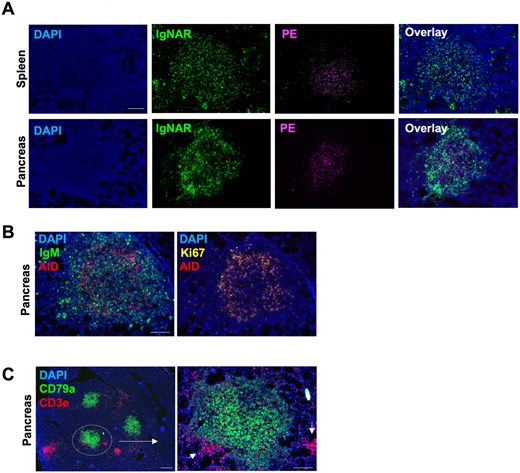
Figure 2.
Pancreatic B cell follicles exhibit functional hallmarks of B cell selection. (A) Fluorescent microscopy images of nurse shark pancreas and spleen tissue showing the localization of nondegraded PE (magenta) in the center of B cell follicles 50 days post-prime with PE. (B) Representative RNAscope in situ hybridization image of nurse shark pancreas showing AID+ and Ki67+ cells localized at the periphery of the B cell follicles. (C) Representative RNAscope in situ hybridization images of nurse shark pancreas show T cell aggregates associated with B cell follicles. Aggregates indicated with arrows. (B–C) Images are representative of 2 nurse sharks that were euthanized 50 d post-prime with PE, providing 2 biological replicates (n = 2). Scale bars = 100 µm.
The presence of non-degraded PE in the center of pancreatic B cell follicles suggested these structures, like those in the spleen, may be environments of B cell proliferation, mutation, and selection. Using RNAscope in situ hybridization, with probes targeting nurse shark AID and Ki67 transcripts, we observed AID signal and Ki67 signal co-localized at the periphery of the pancreatic B cell follicles, suggesting this area is the site of B cell proliferation and BCR mutation (Fig. 2B). Furthermore, cells located at the periphery of the pancreatic B cell follicles were positive for the nurse shark chemokine receptor CXCR4, whereas CXCR5-expressing cells were localized towards the center of the follicles where antigen is located (Fig. S2B). We propose that much like mammalian germinal centers, CXCR4 expression allows AID+Ki67+ B cells to remain on the periphery of the shark pancreatic (and splenic) B cell follicles. Upregulation of CXCR5 and downregulation of CXCR4 would drive these B cells to traffic to the center of the follicles where they test their mutated B cell receptors against the antigen present there.
If pancreatic B cell follicles serve as sites of B cell selection, then T cells should be present within the follicles to provide B cell survival signals. Using RNAscope probes targeting nurse shark CD3e, we identified aggregates of T cells at the margins of the pancreatic B cell follicles with individual T cells scattered throughout the follicles (Fig. 2C). A similar T cell distribution was previously observed in the immunized nurse shark spleen by RNAscope, and single nuclear RNA sequencing (snRNA-seq) indicated a subset of shark splenic T cells are Tfh-like cells capable of providing co-stimulation and survival signals to antigen-specific B cells.14,15 Together, our results suggest that pancreatic B cell follicles possess many of the characteristics necessary for B cell selection, as previously identified in nurse shark splenic B cell follicles.14
Nurse shark pancreas is a site of antigen-specific Ig secretion following immunization
While the discovery of organized immune tissue within the nurse shark pancreas is unexpected, the pancreas as a source of antibody has been indicated by several studies.19–21 Due to the physical link between the pancreas and spiral valve, it has also been proposed that antibody may be secreted directly into the spiral valve via pancreatic ducts to supplement the mucosal immune compartment.19,23 To test this hypothesis, we first sought to determine whether the pancreas serves as a site of antibody production. First, we immunoprecipitated nurse shark Igs from biotinylated pancreas lysate and successfully detected the heavy chains of all three shark Ig isotypes (Fig. 3A). The band for IgM heavy chain (∼75 kDa) was the most prominent, whereas IgNAR (∼90 kDa) and IgW (∼150 kDa) bands were of lower intensity, matching the relative transcript levels observed by RNAscope in situ hybridization (Fig. 1D).
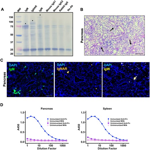
Figure 3.
Nurse shark pancreas is a site of antigen-specific Ig production following immunization. (A) Western blot of biotinylated nurse shark pancreas tissue lysate following immunoprecipitation with antibodies targeting each of the three nurse shark Ig heavy chains or isotype controls corresponding to each primary antibody. Precipitated lysate was run on reducing SDS-PAGE gel prior to transfer. Lane (1) Ladder, (2) Anti-IgM, (3) Anti-IgNAR, (4) Anti-IgW, (5) Anti-Mouse IgG1, (6) Anti-Mouse IgG2, (7) Anti-Rabbit IgG, and (7) no primary antibody. (B) Hematoxylin and eosin staining of nurse shark pancreas tissue sections indicating aggregates of cells around the pancreatic ducts. (C) Representative RNAscope in situ hybridization images of nurse shark pancreas tissue show IgM-, IgNAR-, and IgW-bright cells (indicated with arrows) associated with the pancreatic ducts. (D) Antigen-binding ELISA detects ferritin (Fn)-specific IgM in both pancreas and spleen tissue lysates 100 days post-prime with Fn. Fn-specific IgM binding was compared to that of an unimmunized nurse shark. All lysates were also tested against blocking agent (milk) to assess non-specific binding. Samples were run in duplicate to provide technical replication. (B, C) Images are representative of 2 nurse sharks that were euthanized 50 d post-prime with PE, providing two biological replicates (n = 2). Scale bars = 100 µm.
Initial histological observation of pancreas tissue revealed aggregates of cells surrounding the pancreatic ducts (Fig. 3B). To determine if these cells were B cells and the potential source of Ig, we used RNAscope in situ hybridization to probe for each nurse shark Ig heavy chain isotype. This revealed IgM-, IgNAR-, and IgW-bright cells (ie, putative antigen secreting cells containing very high-levels of Ig transcript) associated with pancreatic ducts (Fig. 3C).
To determine if antigen-specific Ig is produced in the pancreas we immunized two nurse sharks—one with ferritin nanoparticles (Fn) that was euthanized 100-d post-prime, and a second with SARS2 SpFn that was euthanized 50-d post-prime. Pancreas and spleen tissue lysates were then screened for the presence of antigen-specific IgM by ELISA. For the Fn-immunized animal we detected antigen-specific IgM in both the pancreas and spleen lysates with much lower signal observed on blocking agent alone (5% MPBS) and for tissue lysates from an unimmunized animal on either Fn or MPBS (Fig. 3D). Similarly, we detected SARS-CoV-2 RBD-specific IgM in the pancreas and spleen lysates of the SARS-CoV-2 SpFn immunized nurse shark (Fig. S3) with signal above that on MPBS alone. Collectively, these data demonstrate that in conjunction with the spleen, the pancreas contributes to the adaptive immune response in nurse shark by serving as a site of antigen-specific Ig production following immunization.
Nurse shark pancreas houses γδ-enriched T cell clusters that are also positive for AID and Ki67 transcripts
While characterizing the B cell follicles in the pancreas, we also observed large aggregates of CD3e+ T cells that were spatially distant from any CD79a+ B cell aggregates (Fig. 4A). These large clusters of T cells were located throughout the pancreatic tissue and appeared at a similar frequency to the B cell follicles. To our knowledge, such T cell-rich structures have not previously been described in cartilaginous fishes. To further characterize these large T cell clusters, we used RNAscope probes targeting the constant region of the TCR-γ and TCR-α chains. Our probes were first validated on nurse shark thymus tissue, which was positive for both TCR chains with our probes (Fig. S4A); TCR-α and TCR-γ transcript signal was highest in the thymic cortex, where shark T cells undergo somatic recombination through the action of RAG and terminal deoxynucleotidyl transferase (TdT). We also detected AID+ T cells within the thymus (Fig. S4B) as has been observed previously in nurse shark.29 Overall, within the thymus, it appeared TCR-γ+ T cells were in greater abundance than TCR-α+ T cells, which would support previous data indicating that the T cell repertoire is skewed toward γδ T cells in sharks.26
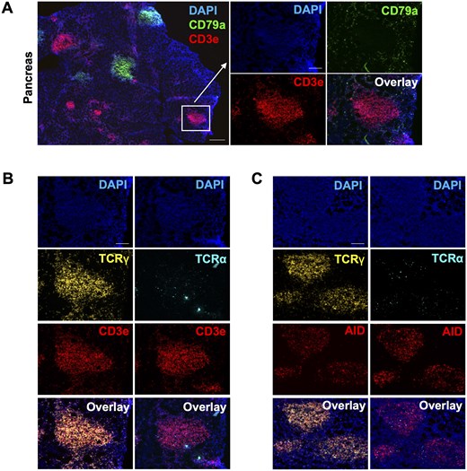
Figure 4.
Nurse shark pancreas houses γδ-enriched T cell clusters that are also positive for AID and Ki67 transcripts. (A) Representative RNAscope in situ hybridization images of nurse shark pancreas show CD3e transcript positive T cell clusters that are spatially distant from the CD79a+ B cell follicles. (B) Representative RNAscope in situ hybridization images of nurse shark pancreas tissue sections show T cell clusters are enriched in γδ T cells. (C) Representative RNAscope in situ hybridization images of nurse shark pancreas show AID transcript expression localized within T cell clusters. (A–C) Images are representative of 2 nurse sharks that were euthanized 50-d post-prime with PE, providing two biological replicates (n = 2). Scale bars = 100 µm.
Using consecutive sectioning of individual pancreatic T cell clusters, we show these clusters are enriched for TCR-γ+ cells with a much lower proportion of TCR-α+ cells (Fig. 4B). This aligns with a previous report that also showed high levels of TCR-γ transcript in nurse shark pancreas with very little TCR-α transcript.26 Surprisingly, we also detected transcripts for AID and Ki67 co-localizing in these pancreatic T cell clusters (Fig. 4C; Fig. S4C). The expression of AID has previously been reported in shark T cells, but only in the thymus, where somatic hypermutation may be utilized for receptor salvaging/editing during positive or negative selection.29 As mutated T cell clones were subsequently identified in the shark spleen, it was suggested that mutated T cells migrate to the periphery after passing tolerance checkpoints in the thymus.30,31 Given we did not observe AID+ T cell clusters in the spleen in this study or in previous work,14 our data demonstrate that the pancreas is a novel niche where T cells may undergo further somatic mutation after leaving the thymus.
Of note, we did not observe the accumulation of intact PE in pancreatic T cell clusters following immunization (Fig. S4D). This would suggest that these T cells are either responding to a processed form of PE presented on MHC (causing it to lose fluorescence) or other unknown antigens. Given the lack of tools for studying shark T cells, we were unfortunately unable to interrogate these T cell clusters any further.
Discussion
While prior reports have suggested that the pancreas of several cartilaginous fish species exhibit immune function,19–21 this finding has never been rigorously investigated. Here, we demonstrate that the nurse shark pancreas houses organized lymphoid tissue that is spatially distinct from the endocrine and exocrine pancreas. This includes pancreatic B cell follicles that exhibit hallmarks of B cell selection as previously found in the nurse shark spleen, including the presence of intact antigen. Furthermore, we also show that the nurse shark pancreas functions as a site of antigen-specific Ig production following immunization.
Importantly, organized B cell follicles were also observed in the pancreas of a healthy, unimmunized control animal, proving that these structures are not an artifact of our immunization regimen but are normal and persistent features within the nurse shark pancreas. Studies of neonatal nurse sharks demonstrate that splenic B cell follicles form early in ontogeny, becoming more “adult like” following dendritic and T cell ingress during the first few months of life.13 Similar studies of fetal and neonatal sharks should shed light upon the development of pancreatic B cell follicles and perhaps uncover if their development is tied to microbial colonization of the gut, analogous to Peyer’s patches in mammals.
Curiously, we also observed organized clusters of T cells that were enriched for TCR-γ, AID, and Ki67 transcripts, suggesting there is a population of rapidly dividing pancreatic γδ T cells that are actively mutating their TCRs. The presence of AID+ Ki67+ T cell clusters in the pancreas generates many questions regarding their contribution to the shark immune response. It is unclear how T cells bearing TCRs mutated in the pancreas would be selected to maintain self-tolerance or MHC compatibility. We did not observe PE in the pancreatic T cell follicles, but the fact that these T cells are dividing suggests they have been activated by antigen. We hypothesize that these T cells, like mammalian γδ T cells, are responding to soluble targets independent of MHC.32,33 Furthermore, given γδ T cells have a conserved role in mucosal protection in other vertebrates34–36 and high levels of γ- and δ-chain transcript have previously been observed in nurse shark mucosal tissues, including the spiral valve,26 we speculate that shark pancreatic γδ T cells diversify their TCR repertoires through SHM before migrating to the spiral valve where they defend the mucosa from pathogens and help maintain gut homeostasis.
While we did not observe major differences in Ig isotype production between the spleen and pancreas, one possible functional distinction could be where the expressed Ig is utilized. Given that we observed an antigen-specific response in the pancreas following subcutaneous immunization, we hypothesize this Ig is secreted directly into the spiral valve via the pancreatic duct, thus protecting the mucosal immune compartment against pathogens encountered in the periphery. Due to technical limitations, we have been unable to test this hypothesis so far. However, we imagine such compartmental crosstalk would be a distinct evolutionary advantage in aquatic organisms that are constantly exposed, inside-and-out, to the same microbe-rich environment. In contrast, terrestrial animals, such as mammals, tend to be exposed to different pathogens via different routes (e.g., skin breach vs oral route) perhaps making a direct link between the peripheral and gastrointestinal compartments, such as that provided by the shark pancreas, less advantageous. Indeed, in humans the B cell clonal responses in the gastrointestinal tract (jejunum, ileum, and colon) are largely distinct from those occurring in other peripheral compartments,37 possibly reflecting differences in the microbiota and pathogens experienced at those sites. However, it should be noted that the human pancreatic B cell repertoire was not examined in the above study, and so the possibility of human pancreas/pancreatic lymph nodes also acting as a “functional bridge” between the two compartments remains unexplored.
Finally, our discovery that the cartilaginous fish spleen, long identified as an SLO,11,13,38 shares this role with another organ is perhaps not as surprising as it first seems. In mammals, it is the coordinated efforts of the spleen, lymph nodes, and GALT that establish timely and effective antibody responses. Also, in at least some teleost fish species the spleen and kidney are both proven SLOs, being able to sample and retain antigen,39,40 while also showing expansion, activation, and SHM of B cells following infection or antigen challenge.41 Indeed, it seems increasingly likely that the sharing of SLO function across multiple sites may be necessary for effective immune surveillance of the periphery and the generation of diverse responses.5,37,42 Prior studies have shown high densities of lymphocyte-like cells and/or significant levels of Ig transcripts in other cartilaginous fish organs, including gut, liver, gonad, brain, gill, and olfactory organ.2,11,13,43–45 Considered alongside the general paucity of information regarding lymphoid tissues in birds, reptiles, and amphibians, we anticipate more SLOs or SLO-like structures await characterization in these lineages. Techniques that enable tracking of antigen and spatial mapping of lineage-determining transcripts/proteins, such as those used here or during the recent discovery of new SLO-like structures in teleost fishes,46,47 will be critical in establishing the organization (or lack thereof) of lymphocytes and other immune cells in these lineages. Knowing the full range of jawed vertebrate organs with SLO potential and how this potential was lost or, more likely, overwritten following the evolution of lymph nodes is key to our understanding of adaptive immune surveillance, humoral protection, and system failure in both non-mammalian and mammalian vertebrates.
Acknowledgments
We thank Dr Mike Crisctiello (Texas A&M University) for sharing the IgWv1 rabbit polyclonal antibody, and Dr Martin Flajnik (University of Maryland, Baltimore) for the NARV and LK14 mouse monoclonal supernatants, used in this study. We thank Dr Alison J. Scott (University of Maryland, Baltimore) for use of her cryostat and for Dr Tialfi Bergamin de Castro (University of Maryland, Baltimore) for sharing his expertise in its use. Thanks to Dr Stuart Weisberg (Columbia University Irving Medical Center) for insightful discussion during the experimental design phase of this work. Also, to Dr Hanover Matz (Washington University) for critical review of our manuscript drafts.
Author contributions
Conceptualization, formal analysis, methodology, visualization, writing—original draft, and writing—review and editing: T.M.H and H.D. Investigation: T.M.H. Resources and supervision: H.D.
Thomas M. Hill (Conceptualization [Equal], Formal analysis [Equal], Investigation [Lead], Visualization [Equal], Writing—original draft [Equal], Writing—review & editing [Equal]) and Helen Dooley (Conceptualization [Equal], Formal analysis [Equal], Methodology [Equal], Resources [Lead], Supervision [Lead], Visualization [Equal], Writing—original draft [Equal], Writing—review & editing [Equal])
Supplementary material
Supplementary material is available at The Journal of Immunology online.
Funding
T.M.H is funded by National Institutes of Health Institutional Training Grant T32AI095190 awarded to Dr Stephanie Vogel (University of Maryland, Baltimore). This work was partly funded by the IMET Innovations in Science Program, sponsored by the G. Unger Vetlesen Foundation, awarded to H.D.
Conflicts of interests
H.D. is a section editor for the Journal of Immunology and has not peer reviewed or made any editorial decisions for this paper. The authors have no financial conflicts of interest.
Data availability
All relevant data are within the manuscript and its supplementary information files.


